
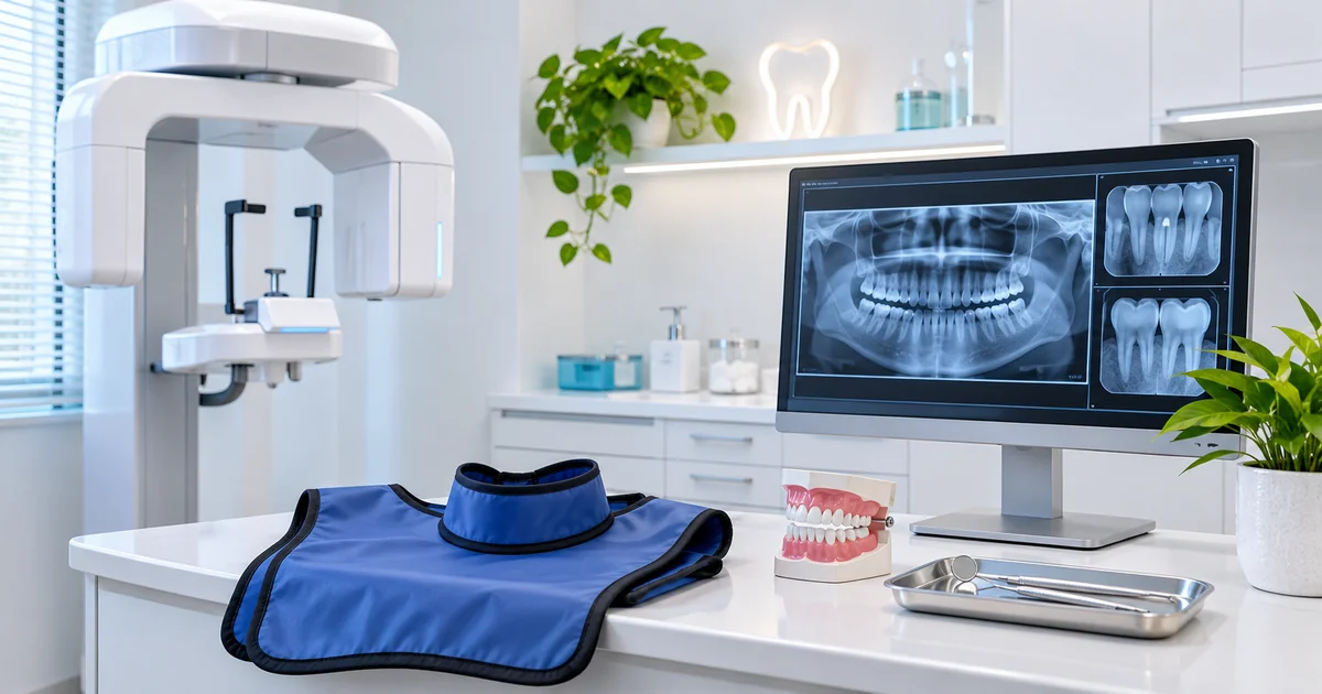
Are dental X-rays safe? Yes, dental X-rays are generally considered safe when used appropriately, with modern imaging techniques designed to keep radiation exposure low. They help dentists identify cavities, infections, bone loss, and other problems that may not be visible during a regular examination. However, X-rays should only be taken when their diagnostic benefits justify the exposure.
At Aesthetic General Dentistry of Frisco, we believe patients should understand the purpose of recommended dental imaging. Knowing how X-rays work, what safety precautions are used, and when images are necessary can help you make informed decisions about your oral health.
Are Dental X-Rays Safe?
Dental X-rays are considered a safe and valuable diagnostic tool when performed for an appropriate clinical reason.
They use a small amount of ionizing radiation to create images of teeth, roots, surrounding bone, and other oral structures.
Although radiation exposure is involved, modern dental imaging techniques are designed to minimize the amount needed to produce useful images.
The American Dental Association (ADA) recommends that dentists order X-rays only when the images are expected to provide information that can improve diagnosis, treatment planning, or patient care.
Important facts about dental X-ray safety include:
- Dental X-rays generally involve low radiation doses.
- Digital imaging can reduce radiation exposure compared with older conventional film techniques.
- Modern equipment can limit the X-ray beam to the area being examined.
- Imaging recommendations depend on individual dental needs.
- Pregnancy does not automatically prevent patients from receiving necessary dental X-rays.
- Unnecessary or repeated images should be avoided.
- Previous dental images may be reviewed before ordering additional X-rays.
Radiation exposure is not completely risk-free, even at low levels.
However, when dental imaging is clinically justified and properly performed, its diagnostic benefits generally outweigh the small associated risks.
The goal is to obtain useful information while keeping radiation exposure as low as reasonably achievable.
How Much Radiation Do Dental X-Rays Use?
One of the most common concerns about dental X-rays is how much radiation patients receive.
Radiation exposure is often expressed in millisieverts (mSv), a unit used to describe effective radiation dose.
Dental X-rays generally produce relatively small doses compared with many other medical imaging procedures.
Understanding dental X-ray radiation levels
The radiation dose depends on several factors, including the type of image, imaging equipment, exposure settings, and number of images taken.
For context, typical reference values published by the United Kingdom’s government include:
| Imaging procedure | Approximate effective radiation dose |
| Single dental bitewing X-ray | Less than 0.01 mSv |
| Panoramic dental X-ray | Around 0.01 mSv |
| Single standard chest X-ray | Around 0.02 mSv |
These figures are examples rather than guaranteed doses for every examination.
Some dental imaging procedures, particularly three-dimensional cone-beam computed tomography (CBCT), may involve higher radiation doses than standard two-dimensional dental X-rays.
How does this compare with everyday radiation?
People are naturally exposed to background radiation from the environment, including the ground, atmosphere, and certain naturally occurring materials.
A single routine dental X-ray often represents a small amount of exposure compared with the radiation people receive from natural sources throughout the year.
However, natural background radiation varies by location, and comparisons do not eliminate the importance of minimizing medical exposure.
Does low radiation mean there is no risk?
Not exactly.
Ionizing radiation can affect biological tissues, and unnecessary exposure should be avoided.
The potential risk from a properly performed routine dental X-ray is considered very small.
Dentists balance this risk against the potential benefit of identifying a dental problem that might otherwise remain undetected.
Why Do Dentists Take Dental X-Rays?
Dentists use X-rays to examine areas that cannot be fully evaluated by looking inside the mouth.
A visual examination provides important information about exposed tooth surfaces, gums, and oral tissues.
However, some dental conditions develop beneath the surface or in areas that are difficult to see.
X-rays help provide additional diagnostic information when needed.
Checking for tooth decay
Certain cavities develop between neighboring teeth or beneath existing restorations.
These areas may not be visible during a routine examination.
Dental X-rays can help dentists identify suspicious changes and determine whether additional evaluation or treatment is necessary.
Evaluating tooth roots
Dental X-rays can show the roots of teeth and surrounding structures.
This is particularly useful when patients experience persistent tooth pain, sensitivity, or symptoms suggesting an infection.
The images may help dentists identify changes near the root that require further investigation.
Examining supporting bone
Dental imaging can help assess bone levels around teeth.
For patients with periodontal disease, certain X-rays may provide information about bone loss and help guide appropriate care.
Planning dental treatment
X-rays may also be useful when evaluating impacted teeth, certain extractions, root canal problems, dental implants, or other treatment needs.
Not every procedure requires the same type of imaging.
Dentists select images based on the information necessary for the individual situation.
What Can Dental X-Rays Detect That an Exam Cannot?
Some dental conditions develop in locations that are difficult or impossible to inspect directly.
X-rays help dentists evaluate these areas, although imaging findings must still be interpreted alongside a clinical examination.
Cavities between teeth
Cavities between tightly spaced teeth can develop without being immediately noticeable.
Bitewing X-rays are commonly used to evaluate these areas.
Detecting decay early may allow dentists to recommend more conservative treatment options.
Problems beneath existing restorations
Dental fillings and crowns can sometimes make it difficult to evaluate certain areas visually.
X-rays may reveal signs of recurrent decay or other changes near dental restorations.
However, not every problem beneath a restoration will appear clearly on an X-ray.
Infection near tooth roots
An infection involving the tissues around a tooth root may create changes visible on dental imaging.
For example, an X-ray might show an area of bone change near the root of a tooth.
The dentist must combine the image with symptoms, clinical testing, and other findings before reaching a diagnosis.
Bone loss associated with gum disease
Periodontal disease can affect the structures supporting teeth.
Dental X-rays can help show changes in bone height and surrounding structures.
This information supports an evaluation of periodontal health.
Impacted or unerupted teeth
Some teeth do not emerge normally into the mouth.
Dental imaging can help dentists determine their position and relationship to nearby structures.
This is often relevant when evaluating wisdom teeth or dental development.
However, X-rays do not reveal every dental problem. Some cracks, early cavities, and soft tissue conditions may require other examination methods.
How Often Do You Need Dental X-Rays?
There is no universal dental X-ray schedule that applies to every patient.
The appropriate frequency depends on your dental history, current symptoms, cavity risk, gum health, age, and previous imaging results.
The ADA’s updated 2026 recommendations emphasize selecting dental X-rays according to individual clinical needs rather than taking images automatically.
Patients with a low risk of cavities
Patients with healthy teeth, consistent oral hygiene, and no recent history of decay may not need dental X-rays as frequently as people with higher cavity risk.
Your dentist will consider previous images and current findings when determining whether new ones are necessary.
Patients with frequent cavities
Individuals who develop cavities regularly may need more frequent imaging.
For example, a patient with multiple recent cavities might benefit from closer monitoring of areas between teeth.
The timing should reflect the patient’s individual circumstances.
Patients with gum disease
Certain patients with periodontal disease may need imaging to evaluate changes in supporting bone.
The number and type of images depend on the extent of the condition and the information needed.
Children and teenagers
Children’s dental imaging needs depend on tooth development, cavity risk, symptoms, and clinical findings.
Dentists use age-appropriate techniques and exposure settings to minimize radiation.
Children should not automatically receive X-rays simply because they have reached a particular age.
Patients with new symptoms
A patient experiencing unexplained tooth pain, swelling, or dental trauma may need imaging even if their previous X-rays were taken recently.
The decision depends on whether another image could help identify the cause.
Your dentist should explain why an X-ray is recommended and how the results may affect your care.
Are Digital Dental X-Rays Different From Traditional X-Rays?
Digital and traditional dental X-rays serve similar diagnostic purposes, but they capture images differently.
Traditional radiography uses photographic film, while digital systems use electronic sensors or imaging plates.
Both involve X-ray radiation.
How digital dental X-rays work
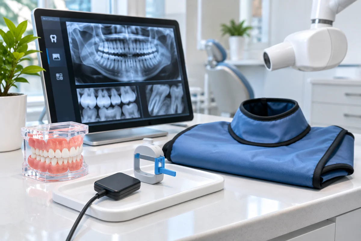
During digital imaging, a sensor or imaging plate captures information as X-rays pass through the area being examined.
The resulting image can be displayed on a computer.
This allows the dental team to review details and, when appropriate, adjust how the image is displayed.
Potential benefits of digital imaging
Digital dental X-rays may offer several advantages:
- Lower radiation exposure: Modern digital receptors can require less radiation than some traditional film systems.
- Faster image availability: Certain systems display images almost immediately.
- Image adjustments: Dentists may enlarge images or adjust contrast to assist evaluation.
- Digital recordkeeping: Images can be stored electronically and compared with previous examinations.
- Easier sharing: Dental records may be transferred securely to another provider when necessary.
However, digital imaging does not completely eliminate radiation exposure.
The actual dose depends on the equipment, imaging settings, and technique.
What about 3D dental imaging?
Three-dimensional imaging, such as CBCT, provides a different type of information from standard dental X-rays.
It may be appropriate for selected complex treatment or diagnostic situations.
Because CBCT can involve greater radiation exposure, it should generally be used only when lower-exposure imaging options cannot provide the necessary information.
What Safety Measures Are Used During Dental X-Rays?
Dental professionals follow radiation safety principles to reduce unnecessary exposure while obtaining useful diagnostic images.
One important principle is ALARA, which stands for As Low As Reasonably Achievable.
This means using the lowest reasonable radiation exposure needed to obtain clinically useful information.
Selecting X-rays only when needed
One of the most effective safety measures is avoiding unnecessary images.
A dentist should consider the patient’s symptoms, dental history, clinical examination, and available previous images before ordering new X-rays.
Limiting the X-ray beam
Modern imaging equipment can restrict the radiation beam to the area being examined.
This is called collimation.
Proper beam restriction helps reduce exposure to surrounding tissues.
Using appropriate exposure settings
Dental imaging settings should reflect the patient and the examination being performed.
Children, for example, require appropriate exposure adjustments rather than automatically receiving adult settings.
Positioning patients carefully
Correct positioning helps produce a usable image the first time.
Poor positioning may require an additional image, resulting in avoidable radiation exposure.
Are lead aprons still necessary?
You may remember wearing a lead apron or thyroid collar during previous dental X-rays.
The ADA updated its guidance in 2024 and no longer recommends routine use of abdominal lead aprons or thyroid collars for dental imaging, including for pregnant patients.
Research indicates that appropriate imaging techniques, beam restriction, and modern equipment provide more effective protection.
Protective shielding can also interfere with the image and occasionally require an additional exposure.
Some practices may still follow specific state or local requirements.
Patients can ask their dental team which safety procedures apply to their examination.
Are Dental X-Rays Safe During Pregnancy?
Yes. When clinically necessary and properly performed, dental X-rays are considered safe during pregnancy.
The American Dental Association states that dental radiographs can be performed during any stage of pregnancy when needed for appropriate dental care.
Why dental imaging may be necessary
Pregnancy does not prevent patients from developing dental problems.
A pregnant patient may experience tooth pain, swelling, or symptoms of an infection requiring evaluation.
If an X-ray is needed to diagnose the problem or guide treatment, postponing necessary care may create additional health concerns.
Does radiation reach the developing baby?
Dental X-rays are directed toward the teeth and surrounding oral structures rather than the abdomen.
With appropriately performed dental imaging, fetal radiation exposure is extremely low.
The evidence supports the safety of clinically justified dental radiographs during pregnancy.
Do pregnant patients need lead aprons?
Current ADA guidance does not recommend routine abdominal or thyroid shielding during dental imaging, including for pregnant patients.
Modern imaging practices focus on restricting the beam and minimizing unnecessary exposure.
However, dental teams must also follow applicable local regulations.
Should you tell your dentist if you are pregnant?
Yes.
Inform your dental team if you are pregnant, believe you might be pregnant, or have relevant medical concerns.
This information helps the dentist coordinate appropriate care and explain why any recommended imaging is necessary.
Pregnancy alone is not a reason to avoid essential dental diagnosis or treatment.
Can You Have Too Many Dental X-Rays?
Although individual dental X-rays generally involve low radiation doses, repeated imaging without a clear purpose should be avoided.
Radiation exposure accumulates from multiple sources throughout life.
This is why dentists should request imaging only when the expected diagnostic benefit justifies the exposure.
What if you recently had dental X-rays?
If you are visiting a new dental office, let the dental team know whether another provider has taken recent images.
Your dentist may be able to review existing records rather than taking additional X-rays.
However, new images may still be needed if the previous ones do not show the area of concern or are no longer sufficient for diagnosis.
Can repeated X-rays increase radiation exposure?
Yes. Each additional examination contributes some radiation exposure.
However, the total amount depends on the type of images taken and their individual doses.
The question is not simply how many X-rays a patient has received, but whether each examination is clinically justified.
Are full-mouth X-rays always necessary?
No.
A full-mouth series may be appropriate for certain patients with extensive dental problems or particular treatment needs.
Other patients may only need selected images.
Routine full-mouth imaging without considering individual circumstances is not consistent with current recommendations for patient-specific imaging.
How Does Your Dentist Decide When X-Rays Are Necessary?
Determining whether dental X-rays are necessary involves reviewing several factors rather than following one fixed schedule.
At Aesthetic General Dentistry of Frisco, we believe patients benefit from understanding the reasoning behind diagnostic recommendations.
Your dental history
Previous cavities, root canal treatments, gum disease, and extensive dental restorations may influence imaging needs.
A history of recurring problems may justify different monitoring than a history of consistently healthy teeth.
Your current symptoms
Persistent sensitivity, pain when biting, unexplained swelling, or dental trauma may indicate that additional imaging would be useful.
Your dentist evaluates whether an X-ray could help clarify the underlying problem.
Your risk of future dental problems
Cavity and gum disease risk can change over time.
Factors such as dry mouth, dietary habits, oral hygiene, existing restorations, and certain medical conditions may affect your needs.
Your previous dental images
Earlier X-rays may contain information that remains useful.
Comparing previous images with current findings can help determine whether additional imaging is necessary.
Whether the results would change treatment
An important consideration is whether the X-ray will provide information that affects diagnosis or treatment planning.
If an image is unlikely to improve the dentist’s understanding or change patient care, it may not be necessary.
Patients should feel comfortable asking questions about the purpose, benefits, and limitations of recommended dental imaging.
Understand Your Dental Imaging Needs at Aesthetic General Dentistry of Frisco
Dental X-rays are one part of understanding your oral health, but they should always serve a clear diagnostic purpose.
At Aesthetic General Dentistry of Frisco, Dr. Chirag Patel and our dental team are committed to helping patients understand their oral health needs and available treatment options.
Whether you have concerns about radiation exposure, questions about previous dental images, or symptoms that may require further evaluation, we encourage you to discuss them with us.
Our approach focuses on explaining recommended care and helping patients make informed decisions based on their individual circumstances.
We welcome patients and families throughout Frisco, TX, who are looking for personalized general and family dental care.
Contact Aesthetic General Dentistry of Frisco at (214) 705-6600 to schedule an appointment and discuss your dental imaging questions with our team.
FAQs
Are dental X-rays safe for children?
Yes. Dental X-rays are generally safe for children when clinically necessary. Dentists use appropriate equipment and exposure settings based on the child’s size and needs while avoiding unnecessary imaging.
Do dental X-rays expose you to a lot of radiation?
No. Routine dental X-rays generally involve low radiation doses. However, exposure varies by examination type, equipment, and number of images, so dentists should only recommend imaging when needed.
Can dental X-rays cause cancer?
Ionizing radiation carries a small potential risk, but the additional risk from an appropriately performed routine dental X-ray is considered extremely low. Avoiding unnecessary imaging helps minimize cumulative exposure.
Are dental X-rays safe during pregnancy?
Yes. Clinically necessary dental X-rays are considered safe during pregnancy. Current ADA guidance does not recommend routine lead shielding, but patients should inform their dentist about their pregnancy.
Do you need dental X-rays every six months?
Not necessarily. Dental X-ray frequency depends on individual cavity risk, dental history, symptoms, age, and clinical findings. There is no universal six-month imaging schedule.
Can a dentist find cavities without X-rays?
Some cavities can be detected during a visual and clinical examination. However, X-rays can help identify decay between teeth or in other areas that are difficult to examine directly.
Are digital dental X-rays safer than traditional X-rays?
Digital dental imaging can use less radiation than older conventional film methods. However, the actual exposure depends on the equipment and technique, and digital X-rays still involve a small amount of ionizing radiation.